
Explain a range of causes of disorders by investigating the structures and functions of the relevant organs, for example:
- Hearing loss
- Visual disorders
- Loss of kidney function
Ears:
- Ears are the organs with which living beings (mammals and birds) hear.
- In case of mammals, ears help in balance as well.
- Structure:
- External or outer ear, consisting of:
- Pinna or auricle. This is the outside part of the ear.
- External auditory canal or tube. This is the tube that connects the outer ear to the inside or middle ear.
- Tympanic membrane (also called the eardrum). The tympanic membrane divides the external ear from the middle ear.
- Middle ear (tympanic cavity), consisting of:
- Ossicles. Three small bones that are connected and transmit the sound waves to the inner ear. The bones are called:
- Malleus
- Incus
- Stapes
- Ossicles. Three small bones that are connected and transmit the sound waves to the inner ear. The bones are called:
- Eustachian tube: A canal that links the middle ear with the back of the nose. The eustachian tube helps to equalize the pressure in the middle ear. Equalized pressure is needed for the proper transfer of sound waves. The eustachian tube is lined with mucous, just like the inside of the nose and throat.
- Inner ear, consisting of:
- Cochlea (contains the nerves for hearing)
- Vestibule (contains receptors for balance)
- Semicircular canals (contain receptors for balance)
- External or outer ear, consisting of:
- Structure:

- Hearing Loss:
- Reduction of hearing ability.
- Inability to hear can be partial or total.
- Most cases, it cannot be cured.
- Causes:
- Advancing age (age-related hearing loss is called presbycusis).
- Certain medications, sometimes called “ototoxic” drugs.
- Trauma or injury to the head.
- Genetic factors.
- Prolonged exposure to excessively loud noise.
- A single episode of acoustic trauma.
- Certain illnesses such as mumps, Meniere’s disease, otosclerosis or autoimmune disease.
- A tumor on the acoustic nerve or acoustic neuroma.
Eyes:
- Eyes provide organisms with the ability to see what is around them and is also passively responsible for a number of photo response that are independent of vision.
- Structure:
- Eye ball consists of three layers
- Outer fibrous layer: Sclera, cornea and conjunctiva
- Middle vascular layer: ciliary body, choroid and iris
- Inner layer: retina
- The eye lens is situated behind iris and consists if the aqueous chamber and the vitreous chamber.
- Eye ball consists of three layers

Common Visual Disorders:
- Myopia:
- Objects nearer to eyes are clearly visible but distant objects are not.
- Occurs when the eyeball is too long, relative to the focusing power of the cornea and lens of the eye.
- This causes light rays to focus at a point in front of the retina, rather than directly on its surface.
- Can also be caused if the cornea and/or lens is too curved for the length of the eyeball.
- Hyperopia / Hypermetropia:
- Distant objects are clearly visible to a person but nearer objects seem blur.
- This occurs when the eyeball is too short, which prevents incoming light from focusing directly on the retina.
- It may also be occurred due to an abnormal shape of the cornea or lens.
- Astigmatism:
- It is a condition in which the eye does not focus light evenly onto the retina resulting into blurry and stretched image formation.
- Occurs when either the front surface of your eye (cornea) or the lens, inside your eye, has mismatched curves. Instead of having one curve.
- May occur in combination with myopia or hyperopia.
Kidneys:
- Pair of organs located in the abdominal cavity that function in removing nitrogenous waste from our body and maintaining homeostasis.
- The structural and functional unit of kidney is nephron which is the centre of all physiological processes carried out in the kidney.
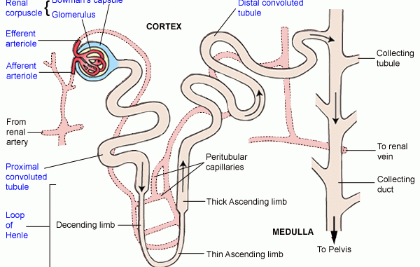
Structure of nephron:
- Nephron is composed of the following parts:
- Renal Corpuscle / Malpighian body (it has two parts)
- Renal Capsule / Bowman’s capsule
- Glomerulus
- Renal tubules (It has 4 parts)
- Proximal Convoluted Tubule (PCT)
- Loop of Henle. It consists of
- Descending limb
- Thin Segment
- Ascending Limb
- Distal Convoluted Tubule (DCT)
- Collecting duct / Collecting tubule
- Renal Corpuscle / Malpighian body (it has two parts)

Loss of kidney function / Kidney Failure:
- Occurs when kidneys lose the ability to filter waste from your blood sufficiently.
- Can be of 5 types:
- Acute prerenal kidney failure
- Caused due to insufficient blood flow to the kidneys.
- The kidneys can’t filter toxins from the blood without enough blood flow.
- Acute intrinsic kidney failure
- Can be caused by direct trauma to the kidneys, such as physical impact or an accident.
- Causes also include toxin overload and ischemia, which is a lack of oxygen to the kidneys.
- Chronic prerenal kidney failure
- When there isn’t enough blood flowing to the kidneys for an extended period of time, the kidneys begin to shrink and lose the ability to function.
- Chronic intrinsic kidney failure
- This happens when there is long-term damage to the kidneys due to intrinsic kidney disease. Intrinsic kidney disease is caused by a direct trauma to the kidneys, such as severe bleeding or a lack of oxygen.
- Chronic post-renal kidney failure.
- A long-term blockage of the urinary tract prevents urination. This causes pressure and eventual kidney damage.
- Acute prerenal kidney failure
Extract from HSC Biology Stage 6 Syllabus. © 2017 Board of Studies NSW.
EasyBio > Non-infectious Disease and Disorders > Technologies and Disorders > Explain a range of causes of disorders
Explain a range of causes of disorders by investigating the structures and functions of the relevant organs, for example:
- hearing loss
- visual disorders
- loss of kidney function
Ears:
- Ears are the organs with which living beings (mammals and birds) hear.
- In case of mammals, ears help in balance as well.
- Structure:
- External or outer ear, consisting of:
- Pinna or auricle. This is the outside part of the ear.
- External auditory canal or tube. This is the tube that connects the outer ear to the inside or middle ear.
- Tympanic membrane (also called the eardrum). The tympanic membrane divides the external ear from the middle ear.
- Middle ear (tympanic cavity), consisting of:
- Ossicles. Three small bones that are connected and transmit the sound waves to the inner ear. The bones are called:
- Malleus
- Incus
- Stapes
- Eustachian tube: A canal that links the middle ear with the back of the nose. The eustachian tube helps to equalize the pressure in the middle ear. Equalized pressure is needed for the proper transfer of sound waves. The eustachian tube is lined with mucous, just like the inside of the nose and throat.
- Inner ear, consisting of:
- Cochlea (contains the nerves for hearing)
- Vestibule (contains receptors for balance)
- Semicircular canals (contain receptors for balance)
- Hearing Loss:
- Reduction of hearing ability.
- Inability to hear can be partial or total.
- Most cases, it cannot be cured.
- Causes:
- Advancing age (age-related hearing loss is called presbycusis).
- Certain medications, sometimes called “ototoxic” drugs.
- Trauma or injury to the head.
- Genetic factors.
- Prolonged exposure to excessively loud noise.
- A single episode of acoustic trauma.
- Certain illnesses such as mumps, Meniere’s disease, otosclerosis or autoimmune disease.
- A tumor on the acoustic nerve or acoustic neuroma.
Eyes:
- Eyes provide organisms with the ability to see what is around them and is also passively responsible for a number of photo response that are independent of vision.
- Structure:
- Eye ball consists of three layers
- Outer fibrous layer: Sclera, cornea and conjunctiva
- Middle vascular layer: ciliary body, choroid and iris
- Inner layer: retina
- The eye lens is situated behind iris and consists if the aqueous chamber and the vitreous chamber.
Common Visual Disorders:
- Myopia:
- Objects nearer to eyes are clearly visible but distant objects are not.
- Occurs when the eyeball is too long, relative to the focusing power of the cornea and lens of the eye.
- This causes light rays to focus at a point in front of the retina, rather than directly on its surface.
- Can also be caused if the cornea and/or lens is too curved for the length of the eyeball.
- Hyperopia / Hypermetropia:
- Distant objects are clearly visible to a person but nearer objects seem blur.
- This occurs when the eyeball is too short, which prevents incoming light from focusing directly on the retina.
- It may also be occurred due to an abnormal shape of the cornea or lens.
- Astigmatism:
- It is a condition in which the eye does not focus light evenly onto the retina resulting into blurry and stretched image formation.
- Occurs when either the front surface of your eye (cornea) or the lens, inside your eye, has mismatched curves. Instead of having one curve.
- May occur in combination with myopia or hyperopia.
Kidneys:
- Pair of organs located in the abdominal cavity that function in removing nitrogenous waste from our body and maintaining homeostasis.
- The structural and functional unit of kidney is nephron which is the centre of all physiological processes carried out in the kidney.
Structure of nephron:
- Nephron is composed of the following parts:
- Renal Corpuscle / Malpighian body (it has two parts)
- Renal Capsule / Bowman’s capsule
- Glomerulus
- Renal tubules (It has 4 parts)
- Proximal Convoluted Tubule (PCT)
- Loop of Henle. It consists of
- Descending limb
- Thin Segment
- Ascending Limb
- Distal Convoluted Tubule (DCT)
- Collecting duct / Collecting tubule
Loss of kidney function / Kidney Failure:
- Occurs when kidneys lose the ability to filter waste from your blood sufficiently.
- Can be of 5 types:
- Acute prerenal kidney failure
- Caused due to insufficient blood flow to the kidneys.
- The kidneys can’t filter toxins from the blood without enough blood flow.
- Acute intrinsic kidney failure
- Can be caused by direct trauma to the kidneys, such as physical impact or an accident.
- Causes also include toxin overload and ischemia, which is a lack of oxygen to the kidneys.
- Chronic prerenal kidney failure
- When there isn’t enough blood flowing to the kidneys for an extended period of time, the kidneys begin to shrink and lose the ability to function.
- Chronic intrinsic kidney failure
- This happens when there is long-term damage to the kidneys due to intrinsic kidney disease. Intrinsic kidney disease is caused by a direct trauma to the kidneys, such as severe bleeding or a lack of oxygen.
- Chronic post-renal kidney failure.
- A long-term blockage of the urinary tract prevents urination. This causes pressure and eventual kidney damage.
Extract from HSC Biology Stage 6 Syllabus. © 2017 Board of Studies NSW.